Tag Archive for: Blog

Breast Cancer Awareness Month: Radiology of Indiana’s Commitment to Comprehensive Care

October is Breast Cancer Awareness Month, a time when we come together to raise awareness, promote early detection, and support those impacted by breast cancer. It’s an important reminder for women to prioritize their health and take proactive steps toward breast cancer prevention and care. Continue reading to learn more about the importance of breast cancer screenings for early detection and some of the advanced imaging options available at Radiology of Indiana.

The Importance of Early Detection

Early detection is one of the most powerful tools in the fight against breast cancer. Studies show that when breast cancer is caught early, the survival rates significantly improve. Regular screening mammograms are essential in identifying abnormalities before symptoms even arise, making it possible to begin treatment at the earliest possible stage. Radiology of Indiana understands that early detection can save lives, and we offer a full spectrum of breast imaging services designed to catch cancer early and provide accurate diagnoses.

Best-in-Class Breast Imaging Services

Radiology of Indiana takes pride in offering state-of-the-art imaging technologies to ensure women receive the highest standard of care. Our board-certified radiologists specialize in breast imaging and work with the latest diagnostic tools to provide precise, thorough results.

Here are some of the key services we offer:

  1. Digital Mammography (3D Mammography): The most common screening tool for detecting breast cancer, 3D mammography allows for more detailed images of the breast tissue, which improves accuracy and reduces the need for follow-up tests. This procedure is particularly beneficial for women with dense breast tissue.
  2. Breast Ultrasound: This imaging technique is often used as a complementary tool to mammography, especially for women with dense breasts. It helps in evaluating abnormalities and provides detailed images of the breast tissue.
  3. Breast MRI: Magnetic Resonance Imaging (MRI) is used in more high-risk patients or to evaluate suspicious areas that are not clearly visible through mammography or ultrasound. Breast MRI is highly sensitive and can detect small tumors that may be missed by other imaging modalities.
  4. Breast Biopsy: If an abnormality is detected, a biopsy may be necessary to determine if the area is cancerous. Radiology of Indiana offers image-guided biopsies, which use mammography, ultrasound, or MRI to precisely locate the area of concern and safely remove tissue samples for evaluation.

Personalized and Compassionate Care

We understand that breast cancer screening and diagnosis can be a stressful experience, which is why our team is committed to providing compassionate, patient-centered care. Our specialists work closely with each patient to guide them through every step of the process, from screening to diagnosis and beyond.

In addition to offering the most advanced imaging techniques, we ensure that every patient feels supported and informed. We collaborate with primary care physicians, oncologists, and surgeons to develop personalized care plans, ensuring that each patient receives the best possible treatment for their unique situation.

Cutting-Edge Treatment Options

For patients diagnosed with breast cancer, access to cutting-edge treatment options is vital. Radiology of Indiana works closely with a network of specialists to provide comprehensive treatment plans. Our advanced imaging technologies assist in planning and guiding surgeries, radiation therapy, and other treatments with precision. We are committed to staying at the forefront of breast cancer treatment advancements, continuously updating our tools and techniques to give patients the highest level of care.

Supporting Breast Cancer Awareness

As part of Breast Cancer Awareness Month, our team encourages women to prioritize their health by scheduling regular mammograms and following recommended screening guidelines. We are proud to partner with the community in spreading awareness about the importance of early detection and offering resources for patients who may need financial assistance or support throughout their breast cancer journey.

Breast Cancer Awareness Month is a reminder of the importance of early detection and high-quality breast care. Our board-certified radiologists provide women with the best-in-class breast imaging services and treatment options. By combining state-of-the-art technology, personalized care, and compassionate support, we aim to make a difference in the fight against breast cancer. Whether you’re coming in for a routine mammogram or need advanced diagnostic services, we are here to ensure you receive the highest standard of care.

Take action this October—schedule your mammogram, spread awareness, and join us in the fight against breast cancer. Learn more at radiologyofindiana.com today.

It’s Time to Tend to Your Tendinitis

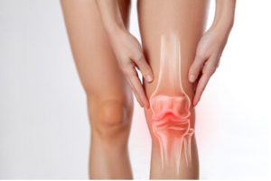
Do you often feel pain or swelling in your muscles and joints? Tendinitis might be the culprit. It’s time to tend to your tendinitis; our latest blog helps to explain the condition further and offers some insight for a deeper understanding. Read to learn more below!

Understanding Tendinitis  

If you’ve ever experienced muscle and joint stiffness, odds are you may have encountered a condition known as tendinitis, the inflammation of a tendon, most often caused by repetitive motion.  It’s a frequent issue that affects any area of the body containing tendons, most commonly near the elbows, wrists, shoulders, and heels. Tendinitis is common in those involved in regular physical activity, such as athletes or manual labor workers who consistently place extra strain on their muscles. It can impact people of all ages alike. Whether it’s a young toddler or a mature adult, the condition has the potential to impact individuals across all stages of life. Pain can range from mild to severe and typically occurs near the affected tendon. Commonly experienced symptoms may include:

  • Pains and aches that are made worse with movement
  • Redness around the inflamed area
  • Reduced flexibility and range of motion
  • Swelling
  • Tenderness

Some individuals may even experience a feeling of heat from the affected area or see a visible lump. As the condition progresses, the pain could intensify, resulting in an inability to perform everyday activities like dressing oneself or something as simple as opening doors. However, early detection and diagnosis can help prevent further worsening of issues and allow time for proper recovery through rest, stretches, and therapy methods. With medical attention and quick action upon signs of discomfort, the condition can be managed before unnecessary problems arise.

Understanding Tendinitis - find out more in our blog!

Tendonitis Treatment Redefined

The options for treating tendon pain could be very limiting, often depending on rest, physical therapy, medications, or open surgery if necessary. As a result, patients often had to wait months or years to gain any relief from their chronic pain. This is changing thanks to the introduction of the Tenex TX procedure. This minimally invasive technique does not require a general anesthetic. Instead the insertion of a Tenex needle into the affected tendon with ultrasound guidance to ensure accuracy. The highly effective treatment removes scarred and damaged tendon tissue and does not require an extended recovery period.

Additionally, by addressing only the affected areas, this procedure can help reduce pain associated with tendinopathy. While also avoiding damage to healthy surrounding tissues. The procedure runs on the short side, only taking about 10-15 minutes, and is often followed with physical therapy.

Interventional radiology - seeing you through to good health! Schedule an appointment today.

Spring has sprung, but alongside it may have come tendon pain. Learn more about how Radiology of Indiana can be of service by learning more about the procedure here or contacting us at (317) 621-5555 today!


Resources:

https://www.hopkinsmedicine.org/health/conditions-and-diseases/tendonitis

https://www.arthritis.org/diseases/tendinitis

https://www.health.harvard.edu/diseases-and-conditions/tendonitis

A Closer Look at Varicose Veins

Varicose veins are a bothersome problem that many of us know little about. This familiar yet undiscussed condition affects one in three adults, yet most patients don’t seek proper treatment. Join us as we take a closer look at varicose veins!

Understanding Varicose Veins – What Are They?

When circulatory system vessels have become abnormally or excessively dilated, it’s known as varicose veins. While more commonly experienced in women, it can affect anyone, with approximately 23% of adults living with it. Varicose veins appear as blue-green twisted or bulging lines beneath the skin’s surface. The veins become enlarged when the valves in the veins no longer function properly and can’t return deoxygenated blood to the heart. As a result, stagnant blood accumulates in portions of the leg veins resulting in pressure buildup and their characteristic swollen appearance. While primarily found in the legs, they can appear elsewhere on the body, such as the feet.

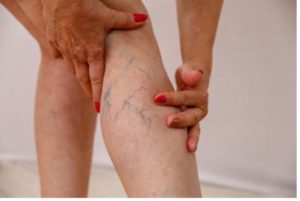
Zoomed in shot of woman with varicose veins.

How to Recognize Varicose Veins

Various factors contribute to the formation of varicose veins—genetics, sedentary lifestyle, extended periods of standing, hormonal changes, and even excessive exposure to the sun. A common myth surrounding varicose veins is that treatment is pursued solely for cosmetic reasons. However, this could not be further from the truth. This condition can cause many downright painful symptoms.

The most common symptoms include:

  • Aching or burning sensations when standing
  • Discoloration around veins
  • Swelling of the ankles and legs

Remember the power of good habits for those fearful of developing varicose veins. Proper care, such as regular exercise, lots of water, weight management, and elevated legs while resting, can minimize your risk for varicose veins. A healthy diet, compression stockings, and the limitation of alcohol are also highly advised.

When to Worry About Varicose Veins

While often harmless, you should seek medical attention if you experience severe symptoms such as intense pain or itching around the veins. Also, look for other signs of complications, such as sores that ooze fluid or bleeding directly from a vein. It’s important to note that varicose veins increase the risk of deep venous thrombosis (DVT) or blood clots. Varicose veins can also decrease mobility and interfere with everyday activities, so it’s vital to remain vigilant of symptoms.

The Role of Interventional Radiology in Treatment

At Radiology of Indiana, we often favor a form of treatment known as Endovenous Laser Ablation due to its efficacy with our patients, with a success rate of approximately 98%. In the past, a surgical procedure known as vein stripping that consisted of entirely removing the vein from the leg was most common. Yet, with the emergence of endovenous laser therapy, surgery is no longer necessary. This speedy procedure will have you walking in and out of our office within an hour, as it does not require general anesthesia. This minimally invasive procedure uses laser energy to seal off problematic varicose veins. By inserting a small laser fiber into a vein through a tiny needle, the laser energy can heat the vein walls and collapse it closed. Covered by all insurance types, treatment options such as endovenous laser ablation are available for those diagnosed with symptomatic varicose veins.

It’s important to note that interventional radiology offers varied treatment options. The best action plan for you can be achieved by scheduling an appointment today. Through radiology, patients can find relief from this painful condition.

Give yourself the care you deserve. Schedule an appointment today.To learn more about interventional radiology services for varicose veins, visit our website or contact us at 317-621-5555 today!


Resources:

https://www.hopkinsmedicine.org/health/conditions-and-diseases/varicose-veins

https://www.nhlbi.nih.gov/health/varicose-veins

https://www.ahajournals.org/doi/10.1161/circulationaha.113.008331#:~:text=Approximately%2023%25%20of%20US%20adults%20have%20varicose%20veins.

Recognizing the Value of Radiologists

Radiologists are some of the most crucial professionals in the medical field but often go unrecognized for their amazing work.  Our latest blog delves into what radiologists do, how they’re making a difference in people’s lives, and recognizing the value of radiologists.

The Role of a Radiologist

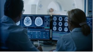
Radiologists celebrate the complexity of the human body and work to improve lives through the use of imaging technology. As medical experts, radiologists are critical in interpreting images captured by radiologic imaging for the diagnosis and treatment of diseases. Primarily used when treating disease and issues in varied areas such as cancer, vascular diseases, men’s health, and women’s health. Through detailed analysis of X-rays, CT scans, ultrasounds, and MRI scans, they can identify injuries and illnesses that could otherwise go undetected. Their skillful work has saved countless lives over the years and will continue to do so in the future.Urologists viewing test results of a patient.

The Relationship Between Radiologist and Patient

 A radiologist plays a key in difficult diagnoses and image-guided treatments of serious medical conditions. Thus, the relationship between radiologists and their patients is unique and one to acknowledge. Working together with other healthcare professionals, they consult patients or their caregivers about the appropriate tests to perform and how to prepare for them.

Additionally, they analyze the images taken and diagnose and explain the results and discuss possible treatments with them. From detecting tumors to uncovering various conditions, they use their expertise and technology to help in providing a diagnosis. They can also provide peace of mind for families who are seeking answers about the health of their loved ones.  Through their expertise, radiologists truly make a difference and help make healthcare diagnoses possible.

We are now hiring for radiologists! Learn more at our careers page.This February, show your body some love. Learn more about all our services as well as explore career opportunities at Radiology of Indiana! Visit our website or contact us at (317) 779-2150 today.

Resources:

https://my.clevelandclinic.org/health/articles/24184-radiologist

https://www.acr.org/Practice-Management-Quality-Informatics/Practice-Toolkit/Patient-Resources/About-Radiology#:~:text=Radiologists%20are%20medical%20doctors%20that,tomography%20(PET)%20and%20ultrasound.

https://www.hopkinsmedicine.org/health/treatment-tests-and-therapies/the-radiologist

8 Possible Signs you May have Fibroids

Uterine fibroids are noncancerous tumors that form in the uterus. However, detection can oftentimes be rather tricky as symptoms are not obvious, if even present at all. Though some women may not show any symptoms, it’s vital to be aware of certain indicators. Read our blog for our top 8 possible signs you may have fibroids!

As a woman, you may develop fibroids, also known as leiomyomas or myomas, which can vary in size from tiny seedlings to large masses. They can occur singly or in groups, and they can grow inside the uterine cavity or outside. Typically developing during the childbearing years but can ultimately occur at any age. While fibroids aren’t cancerous, they can still be dangerous and cause some not-always-obvious symptoms.

The 8 common risk factors of fibroids include:

1) Abdominal pain and cramping

Uterine fibroids are a common cause of abdominal pain and cramping in women. While they don’t typically cause notable symptoms, they can lead to pain and cramping, particularly if they are large or located near the intestinal tract. If you have large fibroids, your doctor may recommend surgery to remove them.

Woman clutching abdomen in pain.

2) Back and leg pain

Back and leg pain is a common complaint among women, especially those of childbearing age. Typically, the pain is attributed to the hormonal changes of the menstrual cycle. However, in some cases, fibroids can cause symptoms like back pain or leg pain. This is because they can put pressure on the nerves in the pelvis, which can cause pain to radiate down the legs.

3) Constipation

One less well-known symptom of fibroids is constipation. This is because they press on the bowel, making it difficult for waste to pass through. In some cases, fibroids can also block the fallopian tubes or obstruct the cervix, causing even more problems with bowel movements. However, the implementation of dietary changes, as well as stool softeners or laxatives, could be of help.

4) Difficulty conceiving

Fibroids can block the fallopian tubes, making it difficult for sperm to reach the egg. They can also interfere with implantation or even prevent the embryo from developing properly. While fibroids are not always responsible for difficulty conceiving, they are one potential cause that should be considered by couples who are struggling to conceive.

5) Feeling bloating or fullness of the abdomen

Fibroids can have several physical manifestations, including feeling full or bloated. This is because they can grow to be quite large and in some cases, press on the stomach and intestines. This pressure may result in swelling and a sensation of being satiated, even with little food intake.

6) Heavy or prolonged menstrual bleeding

Why do fibroids cause heavy bleeding? There are a few reasons. First, fibroids can block the cervix, making it difficult for blood to flow out of the uterus. Second, they can distort the uterine lining, preventing it from shedding properly. And finally, fibroids can increase the production of certain hormones, leading to heavier bleeding and even bleeding between periods.

7) Painful intercourse

Fibroids press on the nerves and blood vessels around the uterus, causing pain and other problems. In addition, fibroids can also cause the uterus to contract erratically, which can be painful for both partners during intercourse.  Their location can play a role in discomfort as well.

8)Urinary frequency or urgency

In some cases, fibroids can cause urinary problems. As they can press on the bladder, causing urinary frequency, urgency, and incontinence. If you are experiencing urinary problems or any of the symptoms above, it’s important to talk to your doctor to rule out any other potential causes and determine the best course of treatment.

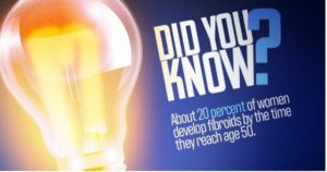
Did you know about 20% of women develop fibroids by the time they reach age 50.

At Radiology of Indiana, we strive to provide top-notch treatment options. We offer fast, non-surgical care to remove fibroids. To learn more, call (317) 621-5555 or visit our website to schedule a consultation today!

Resources:

https://www.radiologyofindiana.com/7-common-symptoms-of-fibroids/

https://www.womenshealth.gov/a-z-topics/uterine-fibroids

https://www.hopkinsmedicine.org/health/conditions-and-diseases/uterine-fibroids

Breast Cancer: The Importance of Imaging

Early detection can save lives; read our latest blog discussing breast cancer and the importance of imaging for detection and possibly treatment!

Breast Cancer Awareness

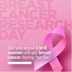
Join us this October as we recognize Breast Cancer Awareness Month because we aim to unite when cancer divides. Breast cancer is a form of cancer that develops in the breast when cells begin to multiply out of control. In the United States, one in eight women will develop breast cancer. Aside from skin cancer, breast cancer is the most common type of cancer among women. In 2022, the American Cancer Society estimates that approximately 43,250 women will die due to breast cancer, with a total of about 287,850 new invasive cases diagnosed.

The two most frequent forms of breast cancer are:

  • Invasive ductal carcinoma:Occurs when abnormal cells form within the milk ducts, then alter and attack breast tissue outside the ducts. Once this occurs, these cancer cells can spread to other body areas. The most common type of breast cancer, making up for a total of 80% of diagnoses.
  • Invasive lobular carcinoma – Beginning in the milk-producing glands known as the breast lobules, as the name invasive implies, this cancer can advance beyond the lobule. It can potentially reach one’s lymph nodes and other parts of the body. This form of breast cancer makes up around 10% of diagnoses.

Early Detection for Your Protection 

When discovered in its early stages, breast cancer has a survival rate of 99%, according to the National Breast Cancer Foundation. Three simple steps can help you remain proactive regarding breast cancer prevention. First, conduct a breast self-examination once a month at home. Familiarize yourself with how they feel and alert your doctor if changes arise. As the saying says, “feel for lumps, save your bumps.” The next step is a clinical breast exam; your physician or gynecologist completes a CBE at your annual examination. They are trained to notice any breast abnormalities or warning signs. The third and final step is a mammogram. This type of imaging allows a specialist to examine the breast tissue of targeted problem areas. Mammograms can detect breast lumps before they can be felt by hand.

Breast Cancer Awareness Pink ribbon.

Early detection is fundamental to treating breast cancer, with varied screening options readily available. Here are different types of radiological imaging used for breast cancer detection:

  • Breast MRI – A breast MRI (magnetic resonance imaging) is a diagnostic exam through a union of radio waves and powerful magnets that forms detailed images of the inside of the breast.
  • Breast Ultrasound – A screening test that utilizes sound waves to look within the breast. Breast ultrasounds also allow for specific breast changes to be monitored, such as a fluid-filled cyst that a mammogram may struggle to depict clearly.
  • Mammograms – Last but certainly not least is the most crucial screening test for breast cancer. Think of a mammogram as an X-ray of the breast, which can detect breast cancer as early as two years before a doctor can physically feel a tumor.

Breast cancer is an extremely difficult disease to experience or watch someone you love the experience. Therefore, raising awareness regarding means of prevention is essential moving forward. As actress and breast cancer survivor Ann Jillian once said, “There can be life after breast cancer. The prerequisite is early detection.”

Breast cancer research day - did you know 1 in 8 women will get breast cancer during her life_

Breast Cancer Awareness is more than just a month. Visit our website today to learn more about breast imaging and the various types provided!

Resources:

https://www.cdc.gov/cancer/dcpc/resources/features/breastcancerawareness/index.htm

https://www.cancer.org/cancer/breast-cancer/screening-tests-and-early-detection.html

https://www.hopkinsmedicine.org/health/conditions-and-diseases/breast-cancer/invasive-ductal-carcinoma-idc#:~:text=What%20is%20invasive%20ductal%20carcinoma,the%20walls%20of%20the%20duct


I changed the image to something a bit more modern. It’s a free image from Pexels!


I think this image was not used so I would find a new one to use from this month’s schedule!


Gotcha!!

Revolutionizing Patient Care with Interventional Radiology

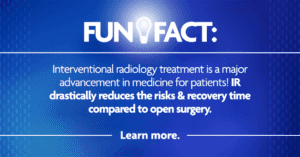
Interventional radiology (IR) is a medical specialty that performs image-guided minimally invasive procedures to diagnose and treat certain conditions. Compared to open surgery, they offer less risk, less pain, and quicker recovery time. With evolving technological advances, revolutionizing patient care with interventional radiology continues to minimize risks and improve patient outcomes.

Experts in Medical Imaging, and More.

Interventional radiology fun fact

Aside from being experts at interpreting medical imaging, interventional radiologists are board-certified physicians with fellowship training in IR procedures. Interventional radiologists use diagnostic images from x-rays, CT scans, MRIs, or other imaging types. The images help them navigate small instruments inside the body to treat various conditions.


The results are often the same or better in comparison to more traditional surgical methods and have other benefits, including:

  • Patient-friendly technology
  • Outpatient or brief hospital stays
  • Tiny incisions minimize scarring
  • Less pain and blood loss
  • Faster recovery time
  • Often less expensive than traditional surgery

Interventional Radiology Procedures

An interventional radiologist works with your provider to treat many of today’s most challenging medical problems, such as cancer, vascular diseases, and men’s and women’s health issues. For example, an interventional radiologist can treat the source of pain or disease by directing a thin wire and a catheter, the size of a strand of spaghetti, through a blood vessel. It may sound unbelievable, but that’s just the tip of the iceberg of the most innovative and effective treatments in medicine occurring today.

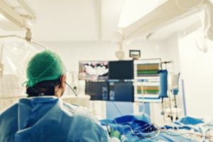
IR procedure

Interventional radiology treatments often have less risk, less pain, shorter recovery times, and better outcomes than traditional (open) surgery. Below are some examples of conditions commonly treated with IR.

  • Cancer
    • Lung
    • Liver
    • Kidney
    • Bone
  • Vascular Diseases
    • Aneurysm
    • Atherosclerosis
    • Blood clots, including deep vein thrombosis and pulmonary embolism
    • Stroke
    • Varicose veins
  • Men’s Health
    • Enlarged prostate
    • Fertility issues
  • Women’s Health
    • Uterine fibroids
    • Chronic pelvic pain
    • Infertility

The Future of Interventional Radiology

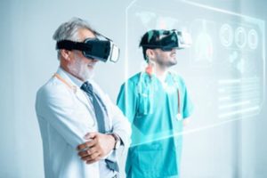
New procedural techniques that include the use of AI, virtual and augmented reality, robotics, and light-sensing technology mean the future of interventional radiology is bright. Since the discovery of the x-ray in 1895, radiology has paved the way for minimally invasive imaging. It is one of the most technologically advanced fields in medicine.

The future of IR

Visit our interventional radiology page at www.radiologyofindiana.com today! You can learn more about the procedures we specialize in, potential benefits, and other frequently asked questions.

Sources:

https://www.diagnosticimaging.com/view/future-trends-and-technologies-in-interventional-radiology-what-to-expect

https://www.sirweb.org/patient-center/

https://www.asrt.org/main/career-center/careers-in-radiologic-technology


Can Bicycling Help Ease Lower Back Pain?

Lower back pain is one of the most common pains experienced universally and can be caused by a variety of things, including injury, improper posture, arthritis, and obesity. However, many have been able to find relief from chronic back pain through exercise. For those looking for a form of physical activity that’s low impact and easy on the joints, cycling may be an excellent option to consider. In this blog, we’ll discuss how, if done properly, bicycling can help ease lower back pain and how to remain mindful of injury when cycling.

Benefits of Bicycling for Low Back Pain

Bicycling is the perfect activity to not only get you outside but also keep your body active. It comes with the added benefit of being low-impact, especially when compared to traditional exercises such as running or weightlifting. Seeing as bicycling puts less overall stress on your body, it can be especially helpful in reducing lower back pain. Bicycling is a form of exercise that doesn’t require much gear and can be done almost anywhere. Additionally, it’s a great option for reducing lower back pain, as it uses a healthy range of motion while providing the core strengthening benefits of an aerobic workout. Bicyclists often say they feel “loose” after a ride. When done correctly, bicycling helps to reduce lumbar muscle tension and improve flexibility—all important contributors to preventing or relieving lower back pain.

Woman exercising at home on cycling machine.

How to Avoid Further Injury When Bicycling

Cycling exercise is a great way to strengthen the lower back when done right. The primary goal should be to maintain good form throughout the ride to ensure proper technique and avoid further injury. To get started, make sure you have an outdoor or exercise bike that fits your body comfortably – handlebars at elbow level, seat position that allows for full leg extension, and back support. Be sure to listen your body. Avoid compromising your posture by vigorously pumping the pedals; instead, look for a steady and smooth rhythm. Keep your spine in a straight line from neck to pelvis, so your lumbar region remains supported while cycling. It’s also important to bring down the intensity – don’t pedal too hard or fast, as this can put strain on your muscles, tendons, ligaments, and discs in the lower back area, which can result in overuse injuries. Physical therapy from cycling can only be beneficial if done properly. With regular practice over time, you can prevent injury when cycling and relieve lower back pain.

Exercise can do wonders when it comes to combating lower back pain relief. Whether decreasing chronic pain, improving flexibility, strengthening muscles around the spine, or reducing stress – the benefits are plenty. By engaging in activities like cycling, you can improve not only your overall physical health but also your mental well-being. Be sure to consult a doctor prior to starting any exercise plan. They will be able to best evaluate how cycling can fit into your individual rehabilitation journey.

Don't let back pain derail you.

There’s nothing to gain from chronic low back pain. Prioritize yourself in 2023 and please visit our website https://www.radiologyofindiana.com/services/back-pain-treatment-choose-your-location/ to find a location to schedule a consultation with one of our Interventional Radiologists.

Resources:

https://www.webmd.com/back-pain/ss/slideshow-exercises#:~:text=Walking%2C%20swimming%2C%20and%20biking%20may,strokes%20that%20twist%20your%20body.

https://cyclingtips.com/2018/10/lower-back-pain-from-cycling-why-it-happens-and-how-to-prevent-it/